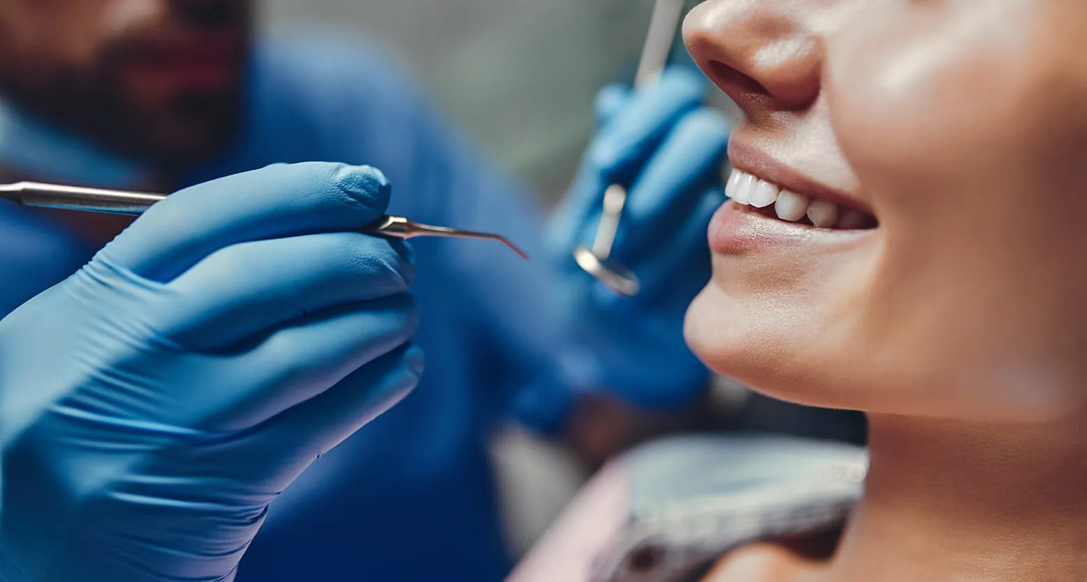
What is an Apicoectomy?
In an apicoectomy, the tip of a tooth root is surgically shortened by a few millimeters, and the surrounding inflamed tissue in the jawbone is removed. Typically, the oral surgeon removes about 2–3 mm of the root tip (the so-called resection) to eliminate the fine branched extensions of the root canal in the apex area, where bacteria often hide, as completely as possible. If necessary, the resected portion may be slightly larger in the case of severely curved roots or other special circumstances.This procedure results in a bacteria-tight seal of the root canal system at the resection site, which enables the healing of a chronic inflammation in the root area. It is important to know that an apicoectomy is not an alternative to root canal treatment, but is usually performed as a supplement—either after an unsuccessful root canal treatment (secondary apicoectomy) or, if no root filling is yet present, in combination with such a procedure during the surgery (primary apicoectomy).
When do we recommend an apicoectomy – reasons and indications for the procedure?
Whether an apicoectomy is necessary depends on the individual findings. Your dentist or oral surgeon will decide, based on clinical examination and X-ray images, whether this tooth-preserving procedure is advisable. Common indications for recommending an apicoectomy include:
- Persistent inflammation: A chronic inflammation at the root tip (apical periodontitis) with persistent pain or a permanent source of infection in the jawbone, which has not healed despite (possibly repeated) root canal treatment. In such cases, the apicoectomy can help remove the source of inflammation directly at the root tip.
- Overfilled root canal material: When root filling material from a previous treatment has gone beyond the root tip into the surrounding tissue and is causing discomfort. Especially if the excess material has entered the sinus cavity or the canal of the mandibular nerve, surgical removal through apicoectomy is indicated.
- Complex root anatomy: Extreme root curvatures or unusual root shapes that do not allow complete cleaning and filling of the root canal by conventional means. In such cases, the apicoectomy can provide access to the root tip from below to remove remaining infected areas.
- Narrowed or blocked root canal: Age-related calcification or obstruction of root canals due to old filling material, which blocks the canals so that they cannot be reached with instruments, even though an infection is present at the root tip. If the root canal can no longer be opened and cleaned from the crown, apicoectomy often remains the only therapeutic option.
- Fractured root canal instrument: A broken instrument lodged in the root canal during a root canal treatment, especially if it is stuck in the area of the root tip and cannot be removed by conventional methods. In such cases, the apicoectomy allows access from below to expose and remove the fragment if it is hindering healing.
- Root perforation: When, during a difficult root canal treatment (e.g., due to severe curvature), a lateral perforation of the root wall near the root tip has occurred. Such an artificial opening can lead to inflammation and usually can only be corrected surgically by resection and secure sealing.
- Root fracture in the apex area: A fracture of the tooth root in the area of the root tip that has led to an infection of the apical fragment. In this case, separating and removing the broken piece (apicoectomy) can be attempted to preserve the remaining tooth.
- Large inflammatory focus (cyst): An extensive inflammatory area of about ≥5 mm in diameter – for example, a radicular cyst at the root tip – may require an additional surgical procedure. In the course of the apicoectomy, the cyst tissue is removed, and the area is carefully cleaned to promote healing.
In general, the necessity for an apicoectomy is always weighed individually based on your specific diagnosis. The procedure serves to preserve the tooth and is usually only performed when less invasive measures (such as another root canal treatment) have been exhausted or are not promising. If an apicoectomy is not feasible or has no prospect of success, the last option often remains tooth extraction followed by tooth replacement (implant or bridge).
Diagnostics & planning: Before an apicoectomy, the situation is thoroughly assessed diagnostically. Based on X-ray images – and, if necessary, also three-dimensionally using Digital Volume Tomography (DVT) – we can precisely assess the extent of the inflammatory focus and the position of the root tip in relation to important neighboring structures (such as the sinus cavity or nerve canal). This 3D diagnostic increases safety and helps plan the procedure optimally to avoid complications.
Procedure: How is an apicoectomy performed?
An apicoectomy is usually performed on an outpatient basis in our practice. Thanks to modern technology and gentle methods, the procedure is generally not very stressful for the patient. The WSR involves the following steps:
- Anesthesia: First, the area to be treated is locally anesthetized. The local anesthesia ensures that the procedure is pain-free. At the patient’s request or in the case of very extensive procedures, sedation in twilight sleep or general anesthesia can also be performed, although the latter is rarely necessary. You must not eat or drink hot beverages while the anesthesia is in effect.
- Exposure of the root tip: After anesthesia, the oral surgeon makes a small incision in the gum in the area of the affected tooth. The gum and periosteum are gently cut and folded back until the jawbone over the root tip is visible. This access from the vestibule (i.e., from the outside of the jaw) allows direct view of the surgical area.
- Bone removal: To reach the root tip, a small amount of jawbone is removed with a special bur. This step is performed very carefully and under constant cooling to protect the surrounding tissue. Once the root tip and the inflamed tissue (such as granuloma or cyst tissue) are exposed, the actual procedure can take place.
- Resection of the root tip: The surgeon shortens the exposed root tip by about 2–3 millimeters. This resection removes the inflamed root tip and the fine branches of the root canal located in the apex area. In some cases, a more extensive shortening is necessary—for example, with severely curved roots or if a metal fragment is lodged in the root tip. The key is to ensure that, at the end of this step, no infected structures remain on the root.
- Cleaning and root filling: The oral surgeon then carefully removes the inflamed tissue from the resulting small bone cavity and rinses/disinfects the area thoroughly. If the tooth did not yet have a root canal filling before the procedure (e.g., because this was not possible due to a blocked canal), the root canal is now opened from the crown of the tooth during surgery, cleaned, dried, and sealed tightly with a root filling. In rare cases, where access from the crown is not possible (such as with an existing non-removable restoration), the canal can alternatively be filled and sealed from the resected root tip (“retrograde”).
- Wound closure: After the root tip is removed and the canal is filled, the surgical site is disinfected again. The surgeon repositions the gum flap and sutures the tissue with fine stitches. This closes the wound so it can heal undisturbed. As a final step, an X-ray is taken to verify the success of the resection and the integrity of the root canal filling. If there was an opening in the crown, the tooth is also given a temporary tight cover to prevent bacteria from entering.
Aftercare and Healing Phase
Proper aftercare plays a key role in ensuring complication-free healing. Immediately after the procedure, you will receive detailed instructions from us. Important: On the day of the surgery, you should avoid certain habits: During the first 24 hours, do not smoke, drink alcohol, coffee, or black tea, and avoid physical exertion. Cooling the cheek from the outside (e.g., with a cold pack wrapped in a cloth) can help relieve and minimize pain and swelling effectively. Additionally, maintain good oral hygiene: Brush your teeth gently and, if necessary, rinse with a suitable antibacterial mouthwash. A clean oral cavity promotes healing and helps prevent wound infections.
In the first few days after the apicoectomy (WSR), slight swelling or throbbing wound pain is normal and usually subsides after a few days. If the pain intensifies or you develop a fever, please contact our practice immediately. The stitches are typically removed after 7–10 days, once the gum at the incision site has healed well. By this time, the soft tissue wound is usually closed. Bone healing takes longer: about 3–6 months after the procedure, a follow-up X-ray is taken to check whether new bone has formed in the area of the removed root tip and whether the inflammation has completely healed.
Risks and Success Prospects of an Apicoectomy (WSR)
As with any surgical procedure, an apicoectomy also carries potential risks. Serious complications are rare, but they should be mentioned:
- General surgical risks: Temporary swelling, bruising, or slight postoperative bleeding can occur. Good cooling and rest usually keep these side effects mild. Wound infections are also possible but occur only rarely if hygiene measures and aftercare instructions are followed.
- Nerve injury: In lower jaw molars, the mandibular nerve runs close to the root tips. In extremely rare cases, this nerve can be irritated or injured during surgery, possibly causing temporary numbness or tingling in the lower lip and chin. Fortunately, permanent nerve damage is very rare.
- Involvement of the maxillary sinus: In upper molars, the root tip is often located near the maxillary sinus. There is a risk that the sinus may be opened during the procedure. If this happens, it is usually treated with special measures (e.g., careful suturing of the sinus membrane window). If left untreated, such an opening could lead to sinus infection, which is why we carefully assess this risk beforehand using CBCT imaging.
- Injury to adjacent teeth: If adjacent tooth roots are located very close together, there is a theoretical risk of damaging the neighboring tooth or its root surface. Through precise planning and gentle operation using magnification aids, we reduce this risk to a minimum.
- Fracture of the remaining root: In rare cases, the remaining tooth root may suffer a crack or fracture during or after the procedure. This can occur if the tooth is already severely weakened or brittle. Such a fracture can worsen the prognosis and, in some cases, may make tooth extraction necessary.
- Persistent or recurrent infection: Although the apicoectomy aims to remove all infected areas, inflammation can sometimes persist or return after some time. Studies show that not every tooth can be saved in the long term — in some cases, the tooth must be removed months or years after the procedure. This often depends on the initial situation (size of the defect, type of bacteria, condition of the tooth). Overall, however, the benefits outweigh the risks: an apicoectomy offers a very good chance of preserving the tooth when otherwise extraction would be necessary.
Despite these possible complications, apicoectomy is a safe and well-proven procedure with high success rates. Modern microsurgical techniques have significantly improved outcomes in recent years. Using magnification aids (operating microscope or surgical loupes) and fine microsurgical instruments allows for extremely precise and tissue-preserving work. This enables complete removal of infected tissue without unnecessarily removing large amounts of bone, which promotes healing. Specialized studies report success rates of over 90% one year after surgery — meaning that more than nine out of ten treated teeth remain symptom-free and functional. Long-term studies in general practice show that around 80–85% of teeth treated with apicoectomy remain healthy for many years. These figures highlight that, in suitable cases, apicoectomy is a very effective tooth-preserving measure.
Modern Technology in Our Practice
As a specialist practice for oral surgery and implantology, we place great importance on providing you with the best possible treatment. For apicoectomies, we rely on modern equipment and gentle techniques:
- 3D X-ray Diagnostics (CBCT): With our digital cone beam computed tomography, we can present the situation in three dimensions. A CBCT scan provides detailed information about the site of inflammation and critical surrounding structures. This allows us to plan the surgery precisely and take potential risks, such as the proximity to nerves or the sinus cavity, into account in advance.
- Microsurgical Equipment: We operate using magnifying loupes or an operating microscope. These magnification aids allow for microsurgical procedures with maximum precision. This enables us to examine and treat the root tip and surrounding tissue in great detail. We also use modern ultrasonic instruments for retrograde root canal preparation, ensuring a tight seal of the canal.
- Sedation and Anesthesia: For anxious patients or very extensive procedures, we offer sedation (twilight sleep) or general anesthesia upon request. In most cases, local anesthesia is sufficient, but with these options, we can make the procedure as pleasant and stress-free for you as possible.
- Experience and Collaboration: As a referral practice for oral surgery, we work closely with your general dentist. We have extensive experience in performing apicoectomies regularly. Our specialization and routine benefit you as a patient: difficult cases (such as multiple roots, large cysts, or old root posts) can be competently assessed and treated. You benefit from short procedure times, a high level of safety, and excellent success rates.
Conclusion: Apicoectomy is a gentle surgical method to preserve your own tooth when deep-seated inflammations cannot be healed otherwise. By combining thorough diagnostics (e.g., with CBCT), modern microsurgical techniques, and our many years of experience, we can, in most cases, preserve the affected tooth for the long term and restore the function and lifespan of your natural tooth. Feel free to contact us – we will provide you with detailed advice and work hand-in-hand with your dentist to find the best solution for you.
Sources: Specialist information and guidelines from the German Society for Oral and Maxillofacial Surgery (DGMKG) and the KZBV patient information were used to create this text, ensuring an accurate and up-to-date presentation of the topic of apicoectomy. All medical details reflect the latest guidelines (AWMF) and are intended to transparently inform you as a patient about both the procedure and the prospects of success of this treatment.
